Summary
Physical Description
Size Range
Colour
Ecology
External Association
Internal Association
Life History & Behaviour
Feeding
Reproduction
Gas Exchange & Excretion
Defence
Anatomy & Physiology
Anatomical Adaptation
Evolution & Systematics
Paleontological Evidence
Biogeographic Distribution
Conservation & Threats
References & Links | 
Comparison of Sarcophyton ehrengergi sclerites abundance between stalk and capitulum
Introduction
The coral reef is known for is amazing biodiversity and bioabundance although nutrients can be low. One of the most important organisms in most reef system is the coral. Corals are important for building substrate as a foundation from their calcium carbonate. However, not all corals have calcium carbonate bodies, like octocorals, specifically soft corals. Instead of building an outer calcium carbonate body, they contain sclerites. Sclerites are a calcareous skeletal component located in the soft coral’s mesogloea (Rao & Devi, 2003). They are formed by fibrous crystals that aggregate and fuse by nanoparticles that are precipitated in the gelatinous network in the mesogloea (Sethmann et al., 2007). Sclerites serve a few purposes in the soft coral, such as structural support as well as defense (Fabricius & Alderslade, 2001). Sclerites play an important role for identifying the species, along side with chemotype and molecular phylogenetic clade identification, due to having various shapes and sizes (Mojaran, 1987; Aratake et al., 2012). It is also important to identify relatedness to sclerites fossils, as the soft bodies of soft corals are difficult to fossilized (Majoran, 1987).
Many studies regarding sclerites have been conducted regarding types of sclerites present in the soft coral species, for example the comparison between the polyp and stalk’s sclerites type (Majoran, 1987; Tentori et al., 2011). However little studies have been done regarding the abundance of sclerites in different location of the soft coral colony, specifically the stalk and the capitulum. The capitulum is rigid and firmer than the stalk due to its need to keep its shape for the polyps to reside in each pore without being crushed, however the stalk is responsible to support the colony as a whole. This study is aimed to investigate the sclerites abundance between the stalk and the capitulum, and is hypothesized that the stalk will have more sclerites compared to the capitulum.
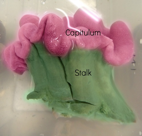
Picture 1 A budding colony from the main colony.
Sarcophyton has been colored to show area of capitulum and stalk
Material & Methods
A small colony was cut off and placed in seawater in a container. Preferably abudding colony as not much fleshy will be used. Subject was cut in half, exposing the inner stalk. A cube piece of 0.5cm 3 sample wastaken from six locations in the stalk as well as the capitulum as shown inpicture 2 and 3.
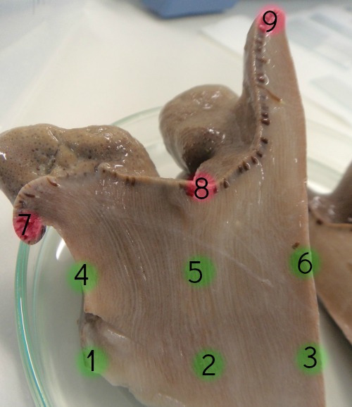
Picture 2 Vertical view of the location where samples were extracted
from the Sarcophyton's stalk and capitulum
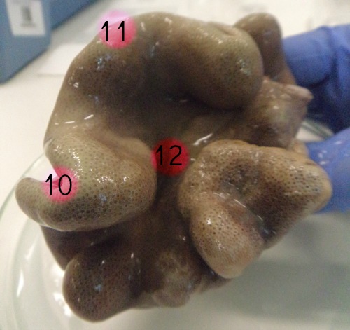
Picture 3 Horizontal view of the Sarcophyton's capitulum of where
the samples were taken from.
The 12 cube pieces are then placed in a well and labeled. Once placed in wells, a scissor was used to cut up the sample into smaller pieces Do not smash the flesh as it was damage the sclerites. Septone bleach was added enough to submerge the sample, and waited for approximately five minutes. Once the flesh has been dissolved, sample was stired and extracted with a pipette. A drop of sample was placed on the slide and sclerites were counter.
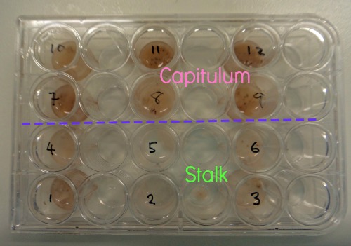
Picture 4 Samples placed in wells and been submerged in septone bleach
Results
Data collected showed higher abundance of sclerites in the stalk collected from the Sarcophyton ehrenbergi (Figure 1). Thus, sclerites are significantly higher in the stalk compared to the capitulum (p<0.05).
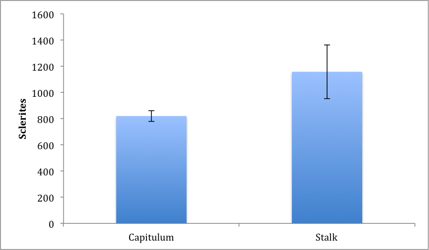
Figure 1 Graph display the average sclerites abundance comparing the capitulum and
stalk samples of Sarcophyton ehrenbergi for six different location each treatment.
Discussion
Results showed that there are more sclerites in the stalk compared with the capitulum. This suggests that sclerites are important for support for the colony as a whole as oppose to providing rigidness to support polyps. It could also suggest that the capitulum’s rigidness is not due to sclerites but some other component, which replaces the sclerites.
Past studies have shown that upper part ofthe stalk had smaller clubbed shaped sclerites with thicker ones towards the basal of the stalk (Majoran, 1987), therefore by having more sclerites locatedat the stalk would give additional support and allow the colony to expand without having the need to develop a calcareous body similar to hard corals.
Although significant results were obtained, it may or may not apply to other colonies with different environmental factors, or a different species. In addition, only a small colony was used, and may differ in larger colonies, and more replicates would be necessary for furtherinvestigation.
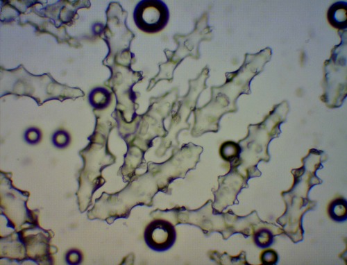 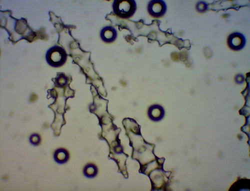 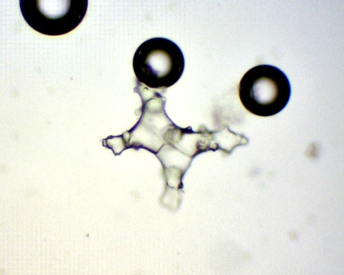
Pic 4 a,b,c Sclerites found from a: basal of the sample, b: capitulum, and c: capitulum.
It can be seen there there are more and thicker sclerites found at the basal compared
to what is found in the capitulum. (c) was found on the capitulum which was not listed
as one of the sclerites for the species Sarcophyton ehrenbergi.
|
|